School program
Concept
ISS2019 is conceived as a unique combination of theory and practical work. The format of the school includes theoretical sessions, where the fundamentals are taught by international top-experts in the field, and hands-on sessions, where theory is applied through practical exercises and workshops. Vendors from leading hard- and software will be invited to the walking dinner, permitting to give the participants a flavour of existing commercial solutions. All lectures will be taught in English.
Informal exchanges during the breaks and lunches offer the possibility to discuss the specific interests of the participants in detail.
Besides the scientific program, social and cultural activities are planned as well (i.e. ice-breaker reception, night tour in Pau, ...).
Program
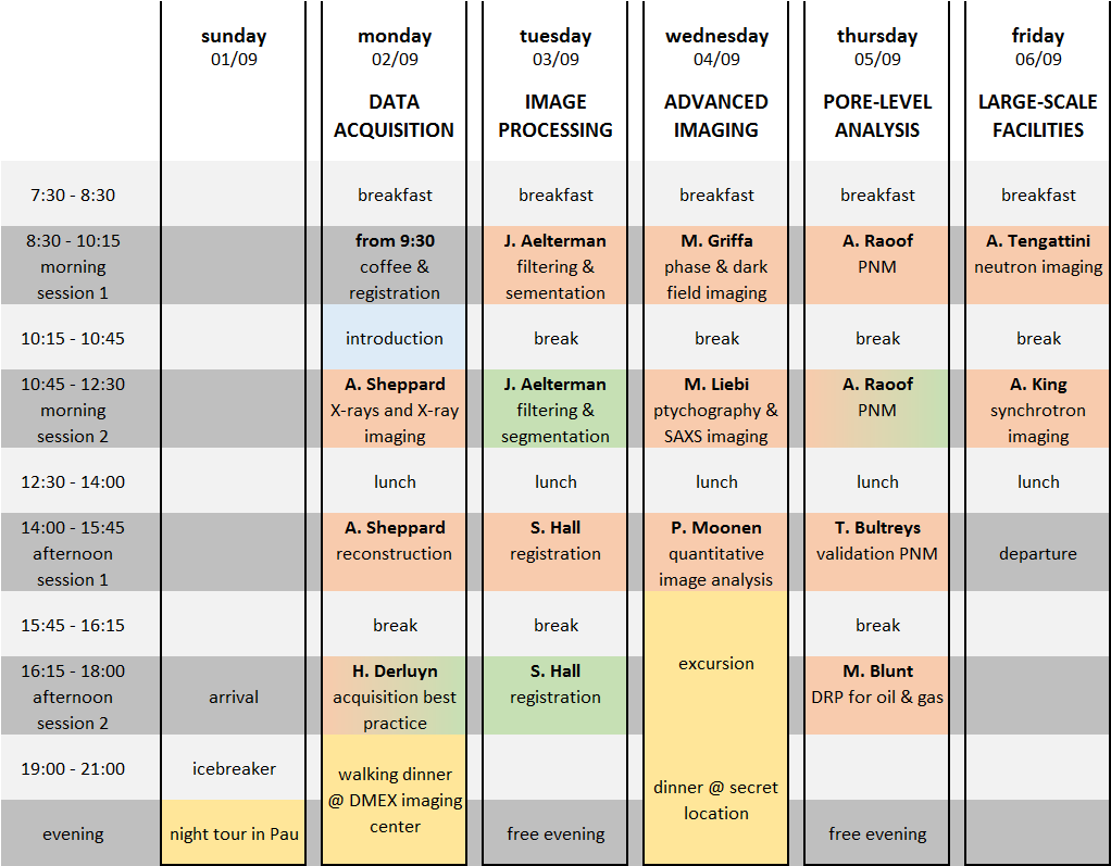
The table below illustrates the tentative school program. Theoretical seminars are indicated in orange and practical work in light green.

Lectures
- X-rays and X-ray Imaging: This talk will cover the fundamentals of X-ray imaging and tomography. Methods for X-ray generation and detection are presented, with an emphasis on laboratory instrumentation, but also covering some more exotic new technologies. I describe the different ways that X-rays interact with matter and the basics of how a 3D tomographic image is constructed from a set of radiographs. I will focus primarily on the factors that limit image resolution, contrast-to-noise ratio and acquisition time, and how to optimise imaging in typical applications.
- Reconstruction methods: This talk will pick up from the morning talk to focus on tomographic image reconstruction. I will cover preprocessing steps and reconstruction using analytical methods, such as filtered backprojection, and iterative reconstruction methods. Reconstruction artefacts caused by system misalignment, beam hardening, phase contrast and sample movement will be discussed, along with methods for ameliorating these artefacts. We will provide a brief introduction to region-of-interest tomography and dynamic (4D) tomography.
- Best practise guidelines: Many parameters affect the quality of an X-ray acquisition. In this lecture best practice guidelines will be given to select optimal parameters, tailored to dynamically visualize pore-scale processes in reservoir rocks.
- Filtering and segmentation: In many fields of science, image analysis involves a scientist setting up an ad-hoc workflow of image processing steps. This lecture will provide insights into the pitfalls and consequences of design decisions in such image processing workflows as well as provide a glimpse into the future of image analysis.
- Registration methods: 4D (3D + time) imaging is a powerful tool to understand processes occurring in materials as they evolve due to, for example, mechanical, chemical, temperature or humidity changes. However, to extract value from 4D image sequences requires appropriate 4D image analysis tools. This lecture will discuss 4D image analysis and, in particular, the use of Digital Image/Volume Correlation techniques to quantify material evolution from time-series of 3D x-ray or neutron images of in-situ experiments.
- Phase and dark-field contrast X-ray imaging: In this lecture I will introduce some basics about two X-ray imaging modalities, namely phase and dark-field con-trast X-ray imaging, respectively, which are becoming increasingly more accessible and widespread and which can provide, in many applications related with imaging liquids in porous media, several advantages compared with the more traditionally used attenuation contrast X-ray imaging.
- Ptychography & SAXS imaging: Besides full field imaging, different types of scanning imaging exist, where a sample is scanned through a focused X-ray beam enabling to record a scattering pattern or fluorescence spectrum in each point. In this lecture SAXS imaging will be introduced, resulting in statistical information on nanostructure in each pixel together with ptychography, where from coherent diffraction high-resolution images can be reconstructed.
- Quantitative image analysis: Grey scales in CT datasets (unfortunately) do not only depend on physical sample quantities such as density and composition. All kinds of scanner (source spectrum, detector efficiency, ...) and scanning protocol (exposure time, ...) related parameters play a role. In this talk we'll analyse some attempts to obtain quantitative data and to assess data uncertainty.
- Pore Network Modelling (PNM): a major application of 3d imaging is its usage for pore scale modeling. In this lecture we will present methods to utilize imaging data for pore scale modeling of two phase flow and reactive transport relevant for oil recovery.
- Validation of PNM: Pore network models try to abstract flow in porous materials by describing the pore space as a network of simple pores and throats, which follow straightforward filling rules. We will show that pore network models can be approached as a tool for analysis and interpretation of pore-scale experiments, and not only as a method for numerical simulations. Using this approach, these models can be validated and augmented by flow experiments imaged with micro-CT.
- Digital Rock Physics for oil&gas applications: The use of pore-scale imaging, analysis and modelling is reviewed with application to the understanding and design of hydrocarbon recovery and carbon dioxide storage. We present recent results that show the fluid/fluid menisci in many natural systems are approximately minimal surfaces – surfaces that minimize area and have an average curvature of zero. This leads to well-connected phases and favourable recovery.
- Neutron Imaging: Neutron follows similar physics to x-rays but interact with the inner nucleus of the atoms, rather then the outer electron shells. This means that the same elements have often completely different attenuation to neutrons than to x-rays, making these two techniques highly complementary. For example Hydrogen and Lithium are almost invisible to x-ray while highly visible to neutrons, while the opposite is true for Aluminium or Quartz . This lecture will provide elements of neutron physics, their production and their usage for high quality imaging with particular focus on hydro-thermo-chemo-mechanical processes in porous media.
- Synchrotron Imaging: I will talk about why we use synchrotron radiation for imaging experiments, how this radiation is produced, and what experiments we can do with it. I will illustrate the talk with examples from experiments performed at the PSICHE beamline.
Activities
- Pau by Night: Guided city walk to discover the rich history of Pau.
- Walking dinner @ DMEX: Visit to the X-ray Imaging center and live demonstrations by Tescan, Voxaya and Zeiss
- Surprise excursion: Half-day excursion to a secret location and dinner at a very original place.


